Atypical Spitz tumour / SAMPUS / MELTUMP
AST · superficial atypical melanocytic proliferations of uncertain significance (SAMPUS) · melanocytic tumour of uncertain malignant potential (MELTUMP)
Atypical Spitz tumour (AST), SAMPUS and MELTUMP are diagnostic categories used by dermatopathologists for melanocytic neoplasms that show atypia exceeding a benign Spitz naevus but do not meet criteria for unequivocal melanoma. They represent a major source of inter-observer variability and clinical uncertainty in dermatopathology. Modern management combines specialist dermatopathology, molecular adjuncts (FISH, CGH, next-generation sequencing), conservative wide local excision and SLNB consideration, with MDT-driven individualised follow-up.
Terminology
- Atypical Spitz tumour (AST): Spitz-like architecture with one or more atypical features (asymmetry, expansile growth, deep mitoses, prominent atypia) — uncertain biological behaviour.
- SAMPUS (Superficial Atypical Melanocytic Proliferations of Uncertain Significance) — confined to epidermis / superficial dermis; doesn't fit clear MIS or naevus categories.
- MELTUMP (Melanocytic Tumour of Uncertain Malignant Potential) — deeper / more atypical; includes atypical Spitz tumour, atypical blue naevus, atypical cellular blue naevus.
- BAP1-inactivated melanocytic tumour (BIMT): separately recognised entity within this spectrum; relevant for BAP1-TPDS.
- The WHO 2023 classification reduces use of these umbrella terms; preferred new framework is molecularly defined (kinase fusions, mutations).
Clinical and pathological context
- Often arises in young patients (paediatric / young adult).
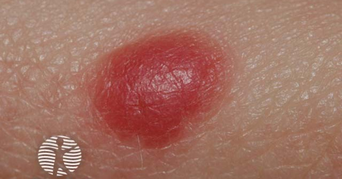
- Pink or pigmented papule / nodule; rapid growth common.
- Sites: face, trunk, extremities.
- Histology shows architectural and cytological features intermediate between benign Spitz / naevus and melanoma.
- Inter-observer variability among expert dermatopathologists is significant — most studies show only moderate agreement.
- Specialist dermatopathology second opinion strongly recommended.
Molecular adjuncts
- FISH panel (4-probe): 6p25 (RREB1), 6q23 (MYB), 11q13 (CCND1), 9p21 (CDKN2A) — gains / losses support malignancy.
- CGH (comparative genomic hybridisation): detects multiple chromosomal aberrations; greater coverage than FISH but less widely available.
- Next-generation sequencing: kinase fusions (ALK, ROS1, NTRK1/3, RET, BRAF, MET, MAP3K8), BAP1 loss, BRAF / NRAS / KIT mutations, TERT promoter mutations.
- Reading the molecular signature:
- Single HRAS mutation alone → favours Spitz.
- TERT promoter mutation → strong adverse prognostic marker.
- Multiple chromosomal gains / losses → support malignancy.
- BAP1 loss → BIMT; consider germline BAP1 testing.
- Increasingly available through tertiary dermatopathology / molecular pathology services.
Management
- Conservative wide local excision: 5-10 mm margins (analogous to MIS) is generally adopted given the diagnostic uncertainty.
- SLNB:
- Considered for atypical Spitzoid tumours with melanoma-like features (Breslow >1 mm, ulceration, high mitotic rate).
- SLN positivity in AST does NOT carry the same prognostic weight as conventional melanoma — multiple studies show SLN-positive AST patients have excellent outcomes.
- Patient counselling about diagnostic uncertainty before SLNB.
- No routine adjuvant therapy — most patients have benign-like outcomes.
- Surveillance: similar to thin melanoma; modified by MDT depending on confidence of diagnosis.
- Multidisciplinary discussion essential:
- Specialist dermatopathology review.
- Molecular pathology adjuncts.
- Discussion with patient about diagnostic uncertainty and management plan.
- Paediatric oncology input for young patients.
- BIMT + suspected BAP1-TPDS: germline BAP1 testing; clinical genetics referral.
Practical points
- Communicate diagnostic uncertainty to patient sensitively but clearly — "not a typical mole; not a definite melanoma."
- Document MDT decision rationale in clinic letter and operation note.
- Photograph the lesion before excision.
- Send specimen for specialist dermatopathology + molecular if uncertain on initial.
- Consider clinical photography / mole-mapping for surveillance.
- Long-term follow-up: tailored to individual histology, molecular profile and MDT consensus.
- Patients with BIMT should be screened for BAP1-TPDS (germline BAP1 testing, MDT input).
References
- Cerroni L et al. Melanocytic tumors of uncertain malignant potential: results of a tutorial held at the XXIX Symposium of the International Society of Dermatopathology. Am J Surg Pathol. 2010;34:314-326.
- Gerami P et al. Fluorescence in situ hybridization for distinguishing nevoid melanomas from mitotically active nevi. Am J Surg Pathol. 2009;33:1783-1788.
- Lallas A et al. Atypical Spitz tumours and sentinel lymph node biopsy: a systematic review. Lancet Oncol. 2014;15:e178-e183.
- WHO Classification of Tumours Editorial Board. Skin Tumours, WHO Classification of Tumours, 5th ed., vol. 12. Lyon: IARC; 2025.
- NICE NG14. Melanoma: assessment and management. London: NICE; 2015 (last updated 27 July 2022).
Spot a correction?
If any clinical statement, citation or link on this page needs updating, please email admin@skinoncology.net with the page name, the proposed correction and the supporting source.