

Aplasia cutis congenita
ACC · congenital absence of skin · congenital scar
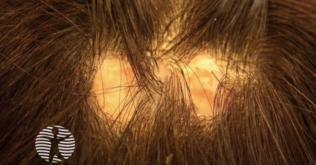
Aplasia cutis congenita is a heterogeneous group of congenital disorders characterised by localised or extensive absence of skin at birth. The commonest form is solitary scalp ACC (Type 1; Frieden classification). Lesions may be simple skin defects, deeper defects involving bone or dura, or part of complex syndromes (Adams-Oliver, trisomy 13, Bart syndrome). Skin-oncology relevance is principally as a clinical sign within Schimmelpenning / naevus sebaceus / Bart syndrome and as a wound-healing / reconstruction challenge.
Frieden 1986 classification
- Type 1: scalp ACC without other anomalies (commonest).
- Type 2: scalp ACC with limb anomalies (Adams-Oliver syndrome).
- Type 3: ACC with epidermal naevi (Schimmelpenning, naevus sebaceus).
- Type 4: ACC overlying embryological malformations (myelomeningocele, omphalocele).
- Type 5: ACC with foetus papyraceus (placental infarction).
- Type 6: ACC with epidermolysis bullosa (Bart syndrome).
- Type 7: ACC limited to limbs (without EB).
- Type 8: ACC caused by teratogens (methimazole, misoprostol, valproate, intrauterine infection — varicella, HSV).
- Type 9: ACC as part of chromosomal / malformation syndromes (trisomy 13, 4p- syndrome).
Clinical features
- Single or multiple well-demarcated areas of absent skin at birth.
- Size: few mm to several cm; may involve bone / dura in deeper lesions.
- Sites:
- Vertex of scalp (commonest — ~70%).
- Trunk, limbs.
- Appearance:
- Glistening atrophic membrane (membranous ACC) — better prognosis.
- Ulcer with granulation tissue.
- Stellate / linear scar at birth.
- Hair collar sign: dense ring of long dark hair around ACC; marker of underlying neural-tube anomaly (cephalocele) → imaging mandatory.
- Frieden Type 2 (Adams-Oliver): limb deficiency (transverse digital anomaly), cutis marmorata telangiectatica congenita (CMTC), congenital heart disease.
- Frieden Type 3: associated epidermal naevus / naevus sebaceus.
Workup
- Imaging:
- Cranial USS or MRI for any scalp ACC with hair-collar sign, bony defect or midline location.
- Spinal MRI if lumbosacral ACC (dysraphism).
- Cardiac echo if Adams-Oliver suspected.
- Examination for limb anomalies (Adams-Oliver), epidermal naevi (Schimmelpenning Type 3), EB phenotype (Bart Type 6).
- Maternal drug / infection history (Type 8).
- Karyotype / microarray if dysmorphic / multi-system.
- Photography for follow-up.
Management
- Small / membranous ACC:
- Conservative — bland emollient, non-adherent dressing.
- Heal by secondary intention over weeks.
- Residual atrophic scar; cosmetic refinement later.
- Large or deep ACC:
- Multidisciplinary plastic / paediatric / neurosurgical input.
- Risk of haemorrhage (especially scalp involving dura / sagittal sinus).
- Risk of meningitis / infection — sterile dressings, prophylactic antibiotics if indicated.
- Surgical: split-thickness skin graft, dermal substitute (Integra), tissue expansion, free flap.
- Syndromic forms:
- Clinical genetics referral.
- Cardiology if Adams-Oliver.
- Surveillance per associated syndrome.
- Long-term cosmetic / reconstructive follow-up; psychological support for family.
References
- Frieden IJ. Aplasia cutis congenita: a clinical review and proposal for classification. J Am Acad Dermatol. 1986;14:646-660.
- Drolet BA, Esterly NB. The hair collar sign: marker for cranial dysraphism. Pediatrics. 1995;96:309-313.
- Bharti G et al. Aplasia cutis congenita: clinical management of a rare congenital anomaly. J Craniofac Surg. 2011;22:159-165.
- Adams FH, Oliver CP. Hereditary deformities in man due to arrested development. J Hered. 1945;36:3-7.
Spot a correction?
If any clinical statement, citation or link on this page needs updating, please email admin@skinoncology.net with the page name, the proposed correction and the supporting source.