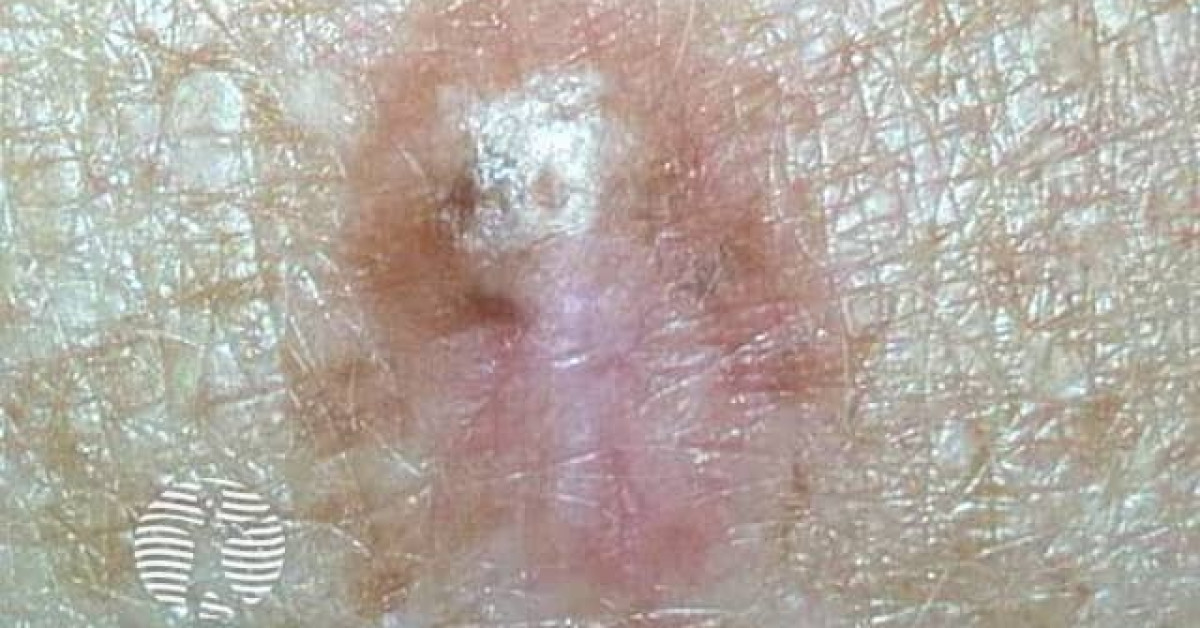
Desmoplastic melanoma
DM; spindle-cell melanoma; neurotropic melanoma (when neural invasion present)
Desmoplastic melanoma is a rare spindle-cell melanoma variant characterised by abundant collagenous stroma and frequent neurotropism. It typically presents on chronically sun-damaged head and neck skin of elderly white patients and is commonly amelanotic, leading to delayed diagnosis. Pure (>90% desmoplastic component) and mixed forms behave differently — pure DM has lower sentinel-lymph-node positivity (<5%) but a higher local recurrence risk, while mixed DM behaves like conventional melanoma. The very high tumour mutation burden makes DM exceptionally responsive to anti-PD-1 immunotherapy.
Epidemiology
- ~2–4% of all cutaneous melanomas; UK incidence increasing in line with population ageing.
- Median age 65–70; M:F ~2:1.
- Strong association with chronic UV damage — head and neck (~50%), upper limb (~30%).
Clinical features
- Slowly enlarging, firm, indurated, often amelanotic plaque or nodule.
- Pigment present in only ~30%; surrounding lentigo maligna in ~30–50%.
- Frequently misdiagnosed as scar, dermatofibroma, BCC or cyst — diagnostic delay is common.
- Neural invasion may produce paraesthesia, dysaesthesia or cranial-nerve palsy when on the face.
Histology & molecular
- Spindled melanocytes embedded in densely collagenous stroma; frequent perineural infiltration ("neurotropism").
- S100 strongly positive; SOX10 positive; HMB-45 and Melan-A often negative — risk of misdiagnosis.
- Pure DM: >90% desmoplastic. Mixed DM: 10–90% desmoplastic.
- Highest tumour mutation burden of any melanoma subtype (UV signature; NF1 loss common; BRAF V600E rare).
Staging & imaging
- Standard AJCC 8 staging applies.
- MRI of the head and neck for facial DM with clinical or histological perineural disease — assess for cranial nerve involvement and skull-base extension.
- SLNB: positivity <5% in pure DM (controversial whether to perform routinely); 15–20% in mixed DM (perform per standard criteria).
Management
- Wide local excision with conventional NICE NG14 thickness-based margins; consider 2 cm minimum and Mohs/peripheral en-face frozen section for facial pure DM given infiltrative growth.
- Adjuvant radiotherapy is used selectively in the UK for pure DM with positive/close margins, perineural disease or head-and-neck sites to reduce local recurrence; the supporting evidence is largely retrospective (e.g. Strom 2014), so use is individualised at the MDT (BAD melanoma 2022 / RCR).
- Advanced/metastatic disease: anti-PD-1 monotherapy (pembrolizumab, nivolumab) — response rates ~70%, far higher than for other melanomas due to the elevated tumour mutational burden.
Prognosis
Local recurrence risk is high (~20–30%) without adjuvant therapy because of infiltrative spindled growth. Distant metastasis tends to be lung-predominant. Overall stage-matched survival is similar to or slightly better than conventional melanoma when local control is achieved. Pure DM has a more favourable nodal profile; mixed DM behaves more like conventional melanoma.
References
- Busam KJ. Desmoplastic melanoma. Clin Lab Med; 2011.
- Eroglu Z et al. High response rate to PD-1 blockade in desmoplastic melanomas. Nature; 2018.
- Strom T et al. Radiotherapy influences local control in patients with desmoplastic melanoma. Cancer; 2014.
Spot a correction?
If any clinical statement, citation or link on this page needs updating, please email admin@skinoncology.net with the page name, the proposed correction and the supporting source.