Halo naevus (Sutton naevus)
Sutton naevus; leukoderma centrifugum acquisitum; halo phenomenon
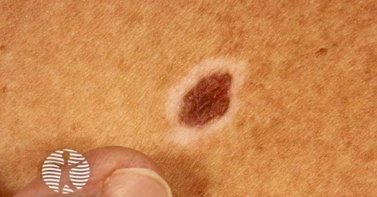
A halo naevus is a benign melanocytic naevus surrounded by a sharply demarcated depigmented halo, representing immune-mediated regression of the central naevus by activated CD8+ T-cells targeting melanocyte differentiation antigens. Multiple lesions are typical, presenting most often in adolescents and young adults on the trunk, back and shoulders. The natural history is one of progressive central naevus pallor (pink → flesh → disappears) over months to years, often followed by gradual repigmentation of the surrounding halo. The condition is benign in essentially all cases. Two clinical considerations matter for skin oncology: multiple halo naevi in an adult may rarely signal an underlying immune response to an occult melanoma (the autoimmune phenomenon being a paraneoplastic marker); and the halo phenomenon is also reported around primary melanomas, so any atypical, asymmetric or rapidly evolving halo lesion requires excisional biopsy.
Clinical features
- Central melanocytic naevus surrounded by a 1–10 mm sharply demarcated depigmented halo.
- Multiple lesions are typical (mean 3–4); occasionally solitary.
- Distribution — trunk, back, shoulders; less often face and limbs.
- Median age 15–25; both sexes; commoner in patients with fair skin.
- Natural history (months to years):
- Stage 1 — central naevus with surrounding halo.
- Stage 2 — central naevus pales pink, then flesh-coloured.
- Stage 3 — central naevus disappears, leaving an achromic patch.
- Stage 4 — gradual repigmentation of the halo.
- Associations — vitiligo (~20% of patients with halo naevi develop vitiligo elsewhere), alopecia areata, Hashimoto's thyroiditis, type 1 diabetes (other autoimmune conditions).
- Painless and asymptomatic.
Dermoscopy
- Symmetrical homogeneous central pigmented naevus structure (network, globules) — depending on stage.
- Sharply demarcated, uniformly white peripheral halo.
- Central regression structures (peppering, white scar-like areas) as the lesion evolves.
- Concerning features — asymmetry, atypical pigment network in the central naevus, multifocal blue-white veil, pseudopods — warrant biopsy.
Differential diagnosis
- Halo phenomenon around melanoma — uncommon but important; primary melanoma can develop a depigmented halo through the same immune mechanism. Asymmetry, atypical pigment, large size, age >40 at first appearance and rapid change should prompt biopsy.
- Vitiligo — depigmented patches without a central naevus; may be associated.
- Post-inflammatory hypopigmentation — irregular borders, no central pigmented lesion.
- Pityriasis alba — children's faces; ill-defined, scaly.
- Naevus depigmentosus — congenital depigmented patch without a central naevus.
Multiple halo naevi in an adult — paraneoplastic concern
- The sudden eruption of multiple new halo naevi in an adult — especially >40 — has been reported as an immune-mediated paraneoplastic marker of an occult melanoma elsewhere on the body.
- Investigation:
- Full skin examination including scalp, mucosa, nails, soles, palms.
- Ophthalmology examination if facial halo lesions or risk factors for uveal melanoma.
- Lymph-node examination.
- If no primary identified — counsel and arrange follow-up; most adult-onset halo naevi remain idiopathic.
Management
- Reassurance — typical halo naevus in an adolescent or young adult requires no treatment.
- Photographic surveillance for any clinically uncertain lesion.
- Excisional biopsy with full histology for:
- Atypical features in the central lesion (asymmetric pigment, irregular border, large size).
- Adult onset (especially >40) of multiple new halo naevi.
- Solitary atypical halo lesion in middle age.
- Diagnostic uncertainty.
- Counsel about photoprotection — depigmented haloes lack melanin and burn easily.
- Counsel about associated vitiligo development.
References
- Mooi WJ, Krausz T. Pathology of melanocytic disorders. 2007.
- Berman B et al. Halo nevus. Cutis; 1979.
Spot a correction?
If any clinical statement, citation or link on this page needs updating, please email admin@skinoncology.net with the page name, the proposed correction and the supporting source.