

Naevus of Ota
Oculodermal melanocytosis; nevus fuscoceruleus ophthalmomaxillaris; naevus of Ito (shoulder distribution)
Naevus of Ota is a congenital or early-acquired dermal melanocytic lesion characterised by a unilateral slate-blue or grey-brown facial discolouration in the distribution of the ophthalmic and maxillary divisions of the trigeminal nerve, with concurrent ipsilateral ocular pigmentation in 60–70%. It is most prevalent in Asian and African populations. Histologically it consists of pigmented dendritic dermal melanocytes — the same lineage as blue naevus — with shared GNAQ/GNA11 driver biology. Although usually benign, naevus of Ota carries an increased lifetime risk of uveal melanoma (and rarely cutaneous melanoma at the affected site or CNS leptomeningeal melanoma), particularly in white patients with the lesion. Dermatology and ophthalmology surveillance is essential, and Q-switched lasers offer excellent cosmetic clearance.
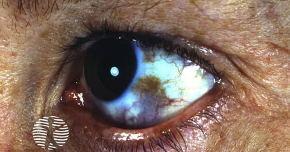
Clinical features
- Unilateral slate-blue, grey or brown discolouration of the periorbital, malar, temporal and forehead skin.
- Distribution corresponds to the V1 and V2 divisions of the trigeminal nerve.
- Ipsilateral ocular involvement (60–70%): blue-grey scleral pigmentation, conjunctival/uveal pigment, iris heterochromia.
- Onset: ~50% present at birth, ~50% during puberty (sometimes hormonally triggered).
- Prevalence: ~1 in 1,000 in Japan; common in East Asian and African populations; rare in white populations but with disproportionate uveal melanoma risk.
- F:M ~5:1.
- Naevus of Ito — same melanocytic process in the supraclavicular, deltoid and scapular distribution.
- Hori's naevus — bilateral acquired equivalent on the malar region of middle-aged women.
Histology & molecular
- Scattered dendritic, pigmented melanocytes throughout the dermis (predominantly upper dermis), with abundant melanophages.
- Identical lineage to the blue naevus family — GNAQ/GNA11 driver mutations.
- Features of malignant transformation include cellular nodules, cytological atypia, mitoses, necrosis, BAP1 loss.
Surveillance & risk
- Uveal melanoma — lifetime risk ~1 in 400 in white patients with naevus of Ota; substantially elevated relative to background. Lower in Asian patients but still increased.
- Cutaneous melanoma at the affected site — rare but reported.
- CNS leptomeningeal melanoma — exceedingly rare; consider in patients with large or extensive lesions or symptoms.
- Ipsilateral open-angle glaucoma — recognised association with ocular involvement; warrants intraocular pressure monitoring.
- Surveillance:
- Annual ophthalmology review with dilated fundoscopy and ocular ultrasound from age 16.
- Intraocular pressure measurement ± gonioscopy for ipsilateral open-angle glaucoma.
- 6-monthly skin examination of the affected and adjacent areas.
- Counsel about melanoma symptoms.
Management
- Treatment is primarily cosmetic and/or for psychological distress.
- Q-switched lasers — Nd:YAG 1064 nm (deeper pigment), Q-switched ruby (694 nm) and Q-switched alexandrite (755 nm) — multiple sessions over 12–18 months yield excellent results. Pico-second lasers may achieve clearance in fewer sessions.
- Camouflage cosmetics for those who decline laser.
- Excisional biopsy of any nodular, atypical or growing focal area.
- Counsel sun protection of the affected skin.
References
- Hidano A et al. Natural history of naevus of Ota. Arch Dermatol; 1967.
- Shields CL et al. Iris and choroidal melanoma in patients with naevus of Ota. Ophthalmology; 2003.
Spot a correction?
If any clinical statement, citation or link on this page needs updating, please email admin@skinoncology.net with the page name, the proposed correction and the supporting source.