Sebaceous carcinoma
Meibomian gland carcinoma; sebaceous gland carcinoma
Sebaceous carcinoma is a rare adnexal malignancy that is most aggressive in periocular sites where it can mimic chronic blepharoconjunctivitis or chalazion. Two anatomical groups — periocular (≈75%) and extra-ocular (≈25%) — differ in clinical behaviour. Suspected cases warrant Mohs micrographic surgery and Muir-Torre syndrome screening.
Clinical features
Periocular
Slowly enlarging yellow-tan or pink eyelid nodule, often on the upper lid (more meibomian glands). Mimics: chalazion, blepharitis, conjunctivitis, BCC. Yellowish hue, eyelash loss (madarosis), and recurrence after chalazion drainage are red flags.
Extra-ocular
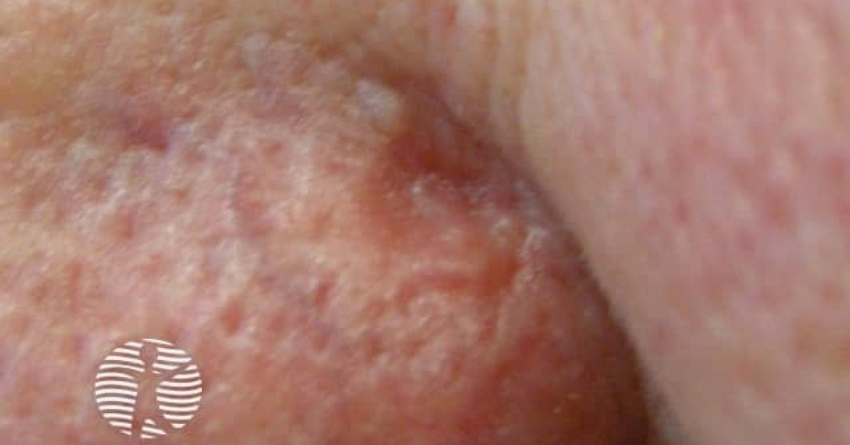
Solitary yellow-pink papule or nodule on head and neck of older adults. Less aggressive than periocular variant.
Diagnosis
Full-thickness eyelid or skin biopsy. Histology: lobules of atypical cells with foamy, vacuolated cytoplasm and high nuclear grade; mitoses prominent. Oil red O on frozen section confirms intracellular lipid; EMA, adipophilin and AR positivity support sebaceous lineage.
Pagetoid spread within epidermis or conjunctival epithelium common.
Muir-Torre screening — tiered MMR algorithm
Tier 1. Tumour immunohistochemistry for MLH1, PMS2, MSH2 and MSH6 (consider EPCAM). Sun-exposed sebaceous tumours frequently show isolated somatic MSH6 loss without a germline cause — MMR loss on a sebaceous tumour does not on its own equate to Lynch syndrome.
Tier 2. Where MLH1 (and consequently PMS2) is lost on IHC, perform MLH1-promoter methylation analysis on the tumour to distinguish the much commoner sporadic cause from Lynch-related germline disease. BRAF V600E testing is the established triage step in colorectal Lynch screening but is less well validated in sebaceous tumours and should be interpreted with caution; methylation is the preferred sebaceous-tumour triage.
Tier 3. Refer to clinical genetics for germline testing where the IHC + methylation triage suggests true Lynch syndrome, where there is a personal or family Lynch-cancer history, or where the patient is young. The Mayo MTS risk score (Roberts 2014) supports pre-test probability where uncertain. See Muir-Torre monograph for the gene-stratified surveillance schedule (Mallorca Group 2022 / NICE NG151).
Management
- Mohs micrographic surgery with frozen-section margin assessment is recommended where available.
- Wide local excision with 5–6 mm margin and paraffin-embedded margin assessment if Mohs unavailable.
- Conjunctival map biopsies for periocular cases to detect pagetoid spread.
- Adjuvant radiotherapy for positive margins where re-excision is not feasible, or for nodal disease.
- Sentinel lymph node biopsy considered for high-risk lesions; metastatic risk approximately 15–20% periocular.
- Immune checkpoint inhibitor (pembrolizumab) — emerging role in advanced / metastatic disease.
Periocular sebaceous carcinoma is best managed jointly by oculoplastic surgery, Mohs / dermatological surgery and the head and neck cancer MDT. Reconstruction often requires staged eyelid reconstruction.
Follow-up
- Clinical surveillance every 6 months for the first 3 years after treatment; continue longer oculoplastic / dermatology follow-up according to stage, ocular involvement, margin status, Muir-Torre / Lynch status and MDT concern.
- Clinical examination of primary site and regional nodes.
- Slit-lamp examination of conjunctiva for periocular disease.
- Lifetime surveillance for Muir-Torre-associated visceral malignancy if applicable.
References
- Owen JL et al. Sebaceous carcinoma: evidence-based clinical practice guidelines. Lancet Oncol; 2019;20:e699–714.
- Kaliki S et al. Sebaceous gland carcinoma of the eyelid. Surv Ophthalmol; 2018;63:457–71.
- Roberts ME et al. A clinical scoring system to identify patients with sebaceous neoplasms at risk for the Muir-Torre variant of Lynch syndrome. Genet Med; 2014;16:711–6.
Spot a correction?
If any clinical statement, citation or link on this page needs updating, please email admin@skinoncology.net with the page name, the proposed correction and the supporting source.