

Drug-induced hyperpigmentation
Drug-induced pigmentation · medication-induced pigmentary disorder
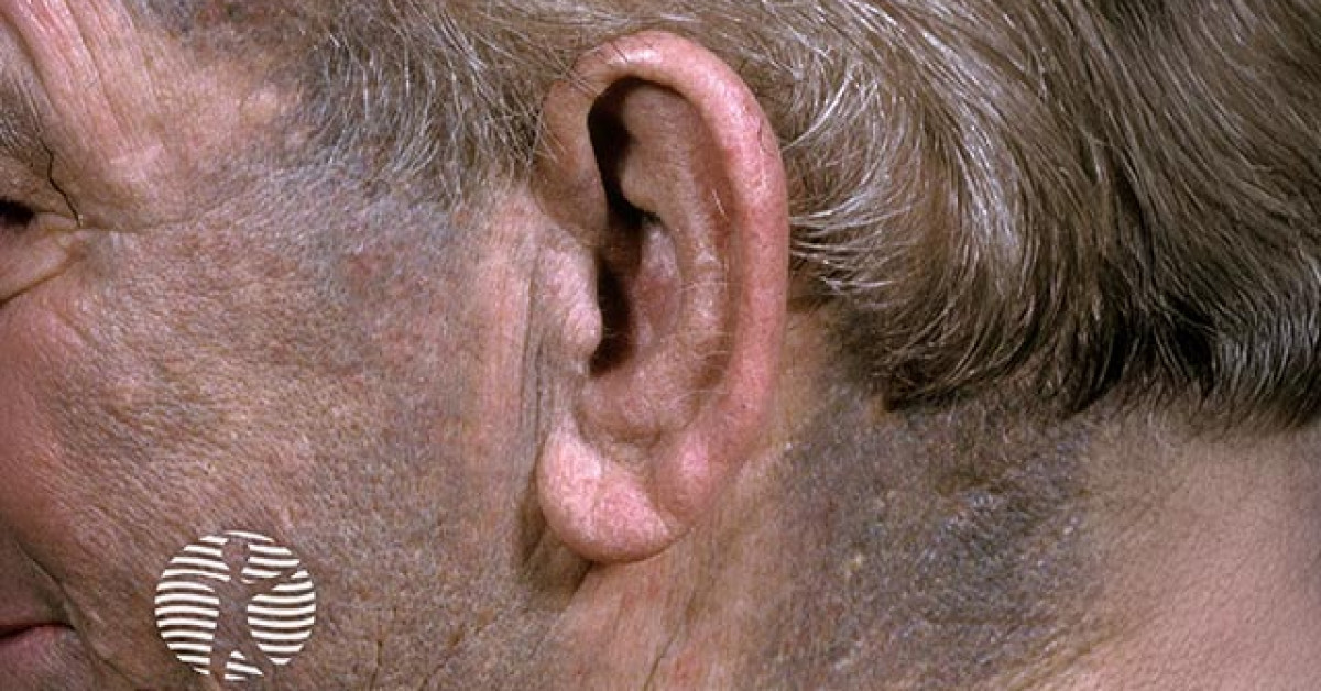
Drug-induced hyperpigmentation accounts for an estimated 10-20% of acquired hyperpigmentation in adults. Several mechanisms are implicated: melanin overproduction, drug-melanin / drug-iron complex deposition, post-inflammatory hyperpigmentation, and direct drug deposition. Skin-oncology relevance: minocycline, antimalarials, chemotherapy (busulfan, bleomycin, cyclophosphamide), EGFR inhibitors, ICI (rare hyperpigmented variant), amiodarone, heavy metals (gold, silver, mercury) — patterns may mimic naevi, melanoma, post-inflammatory change or systemic disease.
Mechanisms
- Melanin stimulation: increased melanocyte activity (oestrogens, phototoxic reactions, ICIs sporadic).
- Direct drug-pigment deposition: minocycline + iron / haemosiderin; amiodarone (lipofuscin).
- Drug-melanin complex: antimalarials (chloroquine, hydroxychloroquine).
- Heavy-metal deposition: silver (argyria), gold (chrysiasis), mercury, bismuth.
- Post-inflammatory hyperpigmentation from preceding drug-eruption / phototoxicity.
Common offenders and patterns
- Minocycline: three types — Type 1 (blue-black in scars / acne sites), Type 2 (blue-grey on shins / forearms / sun-exposed), Type 3 (diffuse muddy-brown on photoexposed skin); years of cumulative exposure.
- Hydroxychloroquine / chloroquine: blue-grey patches on shins / face / mucosae; nail beds.
- Amiodarone: slate-grey / blue photo-distributed on cheeks, dorsal hands; dose-dependent.
- Chemotherapy:
- Bleomycin: flagellate streaks (linear hyperpigmented streaks following scratching).
- Busulfan: diffuse bronzing; mimics Addison disease.
- 5-FU: serpentine supravenous hyperpigmentation along infusion vein.
- Cyclophosphamide, doxorubicin: nail / palmar hyperpigmentation.
- Hydroxyurea: longitudinal melanonychia, palmar / plantar pigmentation.
- EGFR inhibitors: post-inflammatory hyperpigmentation following acneiform eruption.
- BRAF / MEK inhibitors: hyperpigmentation of naevi; reticulate pigmentation; flagellate post-paronychia.
- ICIs: rare vitiligo more common; sporadic hyperpigmentation reports.
- Antiretrovirals: zidovudine — longitudinal melanonychia; mucosal pigmentation.
- Hormonal: oestrogens, OCP — melasma.
- Heavy metals: silver (argyria — slate-grey diffuse); gold (chrysiasis — purple sun-exposed); mercury (perioral / palmar).
Investigations
- Detailed drug history (including supplements, herbal, occupational exposure to heavy metals).
- Wood lamp — distinguishes epidermal from dermal pigment.
- Skin biopsy if uncertain — special stains: Prussian blue (iron), Fontana-Masson (melanin), histochemistry / electron microscopy.
- Atomic absorption spectroscopy for heavy-metal levels (silver, gold, mercury, bismuth).
- Exclude systemic causes — Addison, haemochromatosis, porphyria, jaundice.
Differential diagnosis
- Melasma — symmetric facial; hormonal / UV.
- Post-inflammatory hyperpigmentation — preceding inflammation.
- Lentigo maligna / lentigo simplex — single irregular pigmented patch; dermoscopy / biopsy.
- Naevus, café-au-lait, Becker.
- Addison disease, haemochromatosis, porphyria cutanea tarda.
- Acanthosis nigricans — flexural velvety.
- Erythema dyschromicum perstans (ashy dermatosis).
Management
- Withdraw offending drug where clinically possible — improvement may take months to years; often only partial resolution.
- Photoprotection daily (SPF 50, broad-spectrum) — particularly for amiodarone / chlorpromazine / chemo-related photo-distribution.
- Topical:
- Hydroquinone 4% (intermittent courses), azelaic acid 15-20%, kojic acid, niacinamide.
- Tretinoin / adapalene as adjuncts.
- Triple combination (hydroquinone 4% + tretinoin 0.05% + fluocinolone 0.01%).
- Procedural:
- Q-switched / picosecond lasers (Nd:YAG, alexandrite) for dermal pigment, particularly minocycline, tattoo.
- Caution in Fitzpatrick IV-VI — risk of post-inflammatory hyperpigmentation.
- Counsel that resolution can be slow, especially heavy-metal deposition (often irreversible — chrysiasis, argyria).
References
- Dereure O. Drug-induced skin pigmentation. Am J Clin Dermatol. 2001;2:253-262.
- Krause W. Drug-induced hyperpigmentation: a systematic review. J Dtsch Dermatol Ges. 2013;11:644-651.
- Eisen D, Hakim MD. Minocycline-induced pigmentation: incidence, prevention and management. Drug Saf. 1998;18:431-440.
- Sibaud V et al. Pigmentary disorders induced by anticancer agents. Am J Clin Dermatol. 2018;19:481-498.
- Lerche CM et al. Drug-induced photosensitivity: clinical types of phototoxicity and photoallergy and pathogenetic mechanisms. Front Allergy. 2022;3:838043.
Spot a correction?
If any clinical statement, citation or link on this page needs updating, please email admin@skinoncology.net with the page name, the proposed correction and the supporting source.